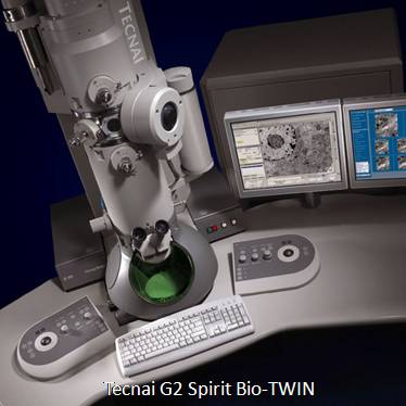
Tecnai G2 Spirit Bio-TWIN Transmission Electron Microscope

  • High contrast, high resolution for 20 –120 kV operation Accelerating Voltage 20, 40, 60 80 100 120 kV
  • Beam source: LaB6 filament.
  • Vacuum system: Oil Diffusion (ODP) and Rotary for projection chamber; Ion (IGP) and Turbo vacuum pumps for column.
  • +/-80˚ fully computer controlled Goniometer stage
  • Optimized for 2D & 3D imaging of cells, cell organelles and soft matter
  • Smart Tracking Position System for sample navigation Low dose mode available
  • Cryo attachment for imaging of biological specimens and materials samples at sub-zero temperatures
  • 4k x 2k Gatan Orius side mount CCD camera.
  • DM3 operation software (Gatan Digital Micrograph)
  • 3D reconstruction software
  • Holders: Standard single tilt; Gatan multiple specimen holder;  Standard single tilt +/- 80˚ Tomography specimen holder; Gatan cryo holder single tilt +/- 80˚ Tomography Cryotransfer Holder

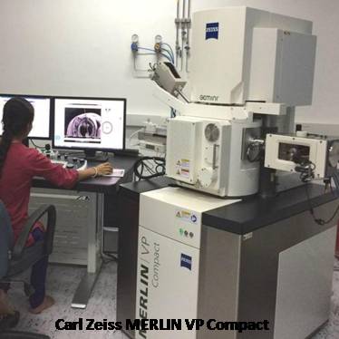
The MERLIN Compact VP

  • Resolution: 0.8nm @ 15KV; 1.4nm @ 1 KV; 0.8nm @ 30KV (STEM mode)
  • Probe current: 5 pA to 100nA
  • Acceleration voltage: 0.02 V to 30 kV 
  • Magnification: 12 – 2,00,000 X
  • Electron Emitter:Thermal (Schottky) field emission gun.
  • Detection Modes
  1. In-lens Duo: Switchable between on-axis in-lens secondary electron detection for highest surface sensitivity and energy selective backscattered detection for advanced materials contrasts.
  2. Secondary Ion: High efficiency in-lens SE detector Everhart Thornley Secondary Electron detector
  3. Variable Pressure: High efficiency VPSE detector for pressure up to 60 Pa
  4. STEM: Low voltage optimized bright field, 4 quadrant dark field, and high angular dark field transmission imaging.
  5. EDAX for chemical composition analysis and element mapping.
  6. Cryo SEM and freeze fracture for imaging hydrated samples or for beam/vacuum sensitive specimens.
  7. Correlative Microscopy with Shuttle and Find.
  • Specimen Stage: 5-Axes Motorized Eucentric Specimen Stage:X = 130mm; Y = 130mm; Z = 50mm; T = - 3º to 70º; R = 360º (continous)  
  • Chamber:  330mm(Ø) x 270mm(h)                              15 accessory ports for various options including CCD-Camera with IR-illumination
  • Vacuum system:  High vacuum mode and variable pressure mode


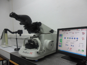
RMC Cryo UltraMicrotome


The Ultramicrotome facilitates preparation of ultrathin sections of biological and polymer material for EM examination. High quality glass knives for ultramicrotomy can easily be fabricated using the Leica Knifemaker in the facility.  Diamond knives are also available for experienced users. There is an cryo attachement available with the RMC ultramicrotome  which enables semi-thin and ultra-thin sectioning of biological specimens and materials samples at sub-zero temperatures.

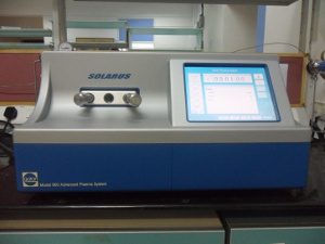
Gatan Plasma Cleaner


Plasma Cleaner enables the removal of impurities and contaminants from surfaces through the use of an energetic plasma created from gaseous species. Gases such as argon , oxygen and hydrogen in different recipes can be used. The plasma is created by using high frequency voltages (typically kHz to >MHz) to ionise the low pressure gas.

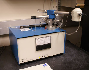
Gatan Carbon Evaporator


Carbon Evaporator can be used for metal sputtering for SEM and TEM applications. It can be used in pulsed or ramped coating modes.

FEI Vitrobot System


Vitrobot is an automated vitrifying equipment for plunge-freezing of aqueous (colloidal) suspensions with a humidity control of 0~100%. The sample can directly be placed on a TEM grid in a humidity and temperature controlled chamber and is then rapidly plunge frozen in liquid ethane preserving the ultrastructure as close as possible to the native state. These samples can then be viewed under the TEM in cryogenic conditions. Applications range from Cryo-EM sample preparation on protein, colloids, virus and bacteria.

Gatan Turbo Pumping Station


This TEM holder module offers intelligent design solutions for cryo-TEM needs and is the perfect compliment to Gatan cryotransfer holders. The self-contain modular bench top pumping station consists of the base unit that contains the vacuum pumps, gauges and electronics and the TEM holder and/or specimen storage modules attached to a specialized vacuum port located on the top surface of the instrument.

Gatan Cryo Transfer Holders


We have set of specimen holders for various applications

  1. Single specimen holder
  2. Multi specimen holder for loading three samples at a time.
  3. Tomography holders with a tilt range of  ±  80°
  4. Cryo Transfer holder.

Sputter Coater

Sputter  coating in SEM is a sputter deposition process to cover a specimen with a thin layer of conducting material, typically a metal, such as gold/paladium. A conductive coating is needed to prevent charging of a specimen with an electron beam in SEM image.

Other Instruments/Accessories

  • Glass Knife cutter
  • Diamond knife
  • The sample preparation room is equipped with two fume hoods, one of them having the air double high efficiency particle arrester (HEPA) as well as carbon filter to maintain clean environment.
  • Benches for the sample preparation have, sputter coater, sonicator, centrifuge, compound and stereo microscopes.
  • Refrigerated storage facility to store heavy metals
  • Oven

 X-ray Micro-Computerized Tomography

  Micro CT Scanner Configuration:
  •    Skyscan 1272, Bruker Instruments
  •    Microfocus sealed X-ray source 20-100kV, 10W, <5um spot size @ 4W.
  •    X-ray detector 16Mp (4904x3280 pixels) or 11Mp (4032x2688 pixels), 14bit cooled CCD fiber-optically coupled to scintillator
  •     Pixel size on the object at maximum magnification [nominal resolution] is 0.35um (16Mp) or 0.45um (11Mp)
  •     Reconstruction arrays – up to 14450x14450x2630pixels (16Mp) or 11840x11840x2150pixels (11Mp) after a single scan
  •     Maximum scanning object diameter -                                                                                                                                                                                                                             1.    75mm (three camera offset positions)                                                                                                                                                                             2.    52mm(two camera offset positions)                                                                                                                                                                                 3.    26mm (central camera position) Fastest Scan
  •    6-position integrated filter changer, standard filters: no filter, Al 0.25mm, Al 0.5mm, Al 1mm, Al 0.5mm + Cu 0.038mm, Cu 0.11mm
  •    Integrated micro-positioning stage, 5-7mm travel (dependent on orientation)
  •    Radiation safety: <1microSv/h in 15cm from any point of instrument surface
 Optional sample changer:
  •     Automatic sample changer with 16 sample positions
  •    Maximum sample size: 25mm diameter, 50 mm length
  •    Sample mounts with four sizes, 16pcs of each size
  •    Scanned samples can be removed / replaced without interrupting scanning process
  •    Typical sample replacement time is 65 sec.
 Control computer:
  •     DELL T7600 Workstation, Windows 7/64bit:     
  •     Two Intel XEON E5-2687W eight-core 2.3GHz processors
  •     128GB 1600MHz RAM
  •     12TB (4x3TB on RAID0) HDD
  •     2GB NVIDIA graphical card + 6GB NVIDIA Tesla C2075 GPU processor
Bruker Image Processing Software:
  •     Skyscan -Scanner and sample changer control, image acquisition
  •     NRecon -Tomography, 3D reconstruction and automatic stitching of oversize scans
  •     CTVox - Volume rendering, pseudo colouration, animation and movie compilation
  •     CTVol -3D surface model editing, 3D structure assembly, animation  and movie compilation
  •     CTAn -Semi-automatic segmentation, 2D/3D  tissue morphometry and surface model generation
  •      Dataviewer - 3D orientation editing, 2D/3D image registration and pseudo colouration
Applications: 
  •    Non-invasive visualization, segmentation and morphometry of insect internal anatomy
  •    Bone density quantification
  •    Surface area, volume, structure thickness, mineral density and pore network analysis in 2D/3D
 

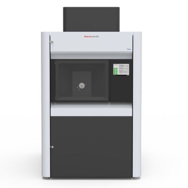
TALOS F200C G2 TRANSMISSION ELECTRON MICROSCOPE

  • Beam Source: X-FEG
  • Accelerating Voltage Range: 80-200kV          
  • +/-60˚ fully computer-controlled Goniometer stage
  • Motorized Apertures
  • Smart Tracking Position System for sample navigation
  • Velox and TIA imaging software's
  • Low dose mode available
  • Holders: Compustage singe tilt holder,
  • Gatan Elsa Cryo transfer holder
  • Gatan 655 Dry Pumping Station for Cryo Holder
  • Ceta-D Camera