
Dr. Mitradas M Panicker - Research
Research Report 2004-2005 :
Gene regulation in the mammmalian nervous system
The major interests of my laboratory are the cellular and molecular regulatory mechanisms active in the mammalian nervous system. We also study events that are involved in early development in mammals, particularly in stem cells.
The interactions of small molecules, such as neurotransmitters with proteins i.e. their receptors expressed on the cell surface play an important role in communication between cells and govern development, behavior, physiology, in fact, all aspects of an organism’s life. One such neurotransmitter is serotonin (5-HT). It is known to play a significant role in depression, stress and sychosis. Among the many receptors that it interacts with, two of these i.e. the 5-HT1A and 5-HT2A receptors have been strongly implicated in stress and in schizophrenia.
Our studies have focused on the regulation of these two receptors in neuronal and non-neuronal cells and also its role in early developmental processes. During stress, corticosteroid levels are significantly affected and lead to decreased levels of the 5-HT1A receptor. Using a model neuronal cell line, generated in our laboratory, we can mimic some of the regulation seen in the mammalian brain. The 5-HT2A receptor, on the other hand, is an important target of many clinically prescribed antipsychotics. Using modified 5-HT2A receptors, which can be visually localized within cells, we have
made significant observations regarding the behavior of the receptor in the presence of serotonin and antipsychotics. These studies should now help us dissect the details of how these receptors are regulated during stressful and abnormal situations.
Our recent results using embryonic stem cells also suggest that some of these receptors and serotonin may play an important role in early differentiation.
1. Effects of agonists and antipsychotics on the internalization of serotonin 2A (5-HT2A) receptor in neuronal and non-neuronal cell lines
Samarjit Bhattacharyya, Aditi Bhattacharya, Saptarshi Mandal and Ishier Raote
5-hydroxytryptamine 2A (5-HT2A) receptors are seven transmembrane G protein-coupled receptors, which are widely distributed in the mammalian nervous system whose natural agonist is serotonin. Depression, suicide, schizophrenia and many other mental and neurological illness seem to be affected by this receptor and its responsiveness to a variety of drugs.
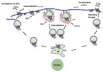
Prolonged exposure of the receptor to ligands is often accompanied by a decrease in response termed desensitization. Internalization of the receptor is also often observed. The phenomena of receptor desensitization and down-regulation are well described in 5-HT2A receptors, although the details of these mechanisms of desensitization are yet to be understood. We have tagged the C-terminus of the full-length 5-HT2A receptor with a number of fluorescent proteins, such as GFP, and have been able to follow the receptor through the process of desensitization and resensitization within neuronal and non-neuronal cells. The receptor is found to internalize on binding to serotonin and some antipsychotics via the clathrin coatmediated pathway and the process is dynamin-dependent. The receptor is coupled to the IP3 pathway and activation of protein kinase C (PKC) is necessary and sufficient to cause the internalization of the 5-HT2A receptor (1). The internalized receptors recycle back to the surface and the kinetics of internalization and recycling of the receptor is faster in neuron-like cell lines (N1E-115 and P40H1) as compared to a non-neuronal cell line (HEK293).
Various typical and atypical antipsychotic drugs act as antagonists on 5-HT2A receptors and some of these cause the receptor to internalize. In general, typical antipsychotics e.g. haloperidol do not cause the receptor to internalize whereas atypical antipsychotics e.g. clozapine, olanzapine do. We have determined that antipsychotic-internalized recycle to the cell surface but take longer time to do so when compared to serotonin-internalized receptors. The internalization is also not dependent on activation of PKC (2). Our current research interests are to identify the molecular mechanisms of agonist and antagonistmediated internalization of the 5-HT2A receptor and its various sub-cellular fates.
2. Role of 5-HT2A receptors in cell adhesion
Basudha Basu
We have observed that expression of the 5-HT2A receptor in HEK-293 cells causes these cells to show increased adhesion to a number of substrates in vitro . The adhesion is decreased in the presence of a number of inverse agonists or antagonists (antipsychotics) of the receptor. We have determined that the transcripts of a number of adhesion-related genes are upregulated in the presence of the activated receptor and antagonists, in turn, cause these gene transcripts to be down-regulated. A similar phenomenon seems to occur in vivo , in a mouse model that we are studying. These studies suggest that antipsychotics may have multiple effects including changes in cellular adhesion.
3. Generation and differentiation of Human and Mouse Embryonic Stem Cells
Imtiaz Zafar and Basudha Basu
Embryonic stem (ES) cells are pluripotent, self-renewing cells derived from the inner cell mass of the developing balstocyst. We have used ES cells as a model system to study neuronal differentiation in vitro . In particular, the roles of serotonin and serotonin receptors in this process are being investigated.
Mouse ES cells have been reported to synthesize serotonin. We have confirmed the presence of serotonin in these cells by antibody staining and multi-photon microscopy. We have shown using RT-PCR, that ES cells also express a subset of serotonin receptors which includes the 5-HT2A receptor. The 5-HT2A receptors in these cells are functional and 5-HT2A transcripts have been detected at various stages of differentiation of ES cells. Blocking 5-HT2A receptor function using antagonists at certain stages of differentiation of
ES cells perturbs the process. Further studies are being carried out to understand the role of serotonin and its receptors in ES cells (3).
We plan to carry out similar studies on human embryonic stem cells. In addition we are also attempting to generate new human embryonic stem cell lines.
Collaborators: Mehroo Hansotia, Sadhana Desai, Vijay Mangoli and Ranjana Mangoli, The Fertility Clinic, Mumbai and Sudipta Maiti, TIFR, Mumbai.
4. Transcriptional regulation of 5-HT1A receptors
M.S. Geetha and Ashok. C
The 5-HT1A receptor is a target for a number of anxiolytics and mice lacking this receptor show increased anxiety. It has also been reported that corticosteroids down-regulate the expression of these receptors in the hippocampus in vivo . P40H1, a neuron-like conditionally immortalized hippocampal cell line generated in the laboratory earlier, expresses 5-HT1A receptors. We observe that in these cells 5-HT1A transcripts are down-regulated in the presence of corticosteroids. Constructs with various segments of the upstream promoter regions of the mouse 5-HT1A gene, that we have designed, also show differential regulation in these cells. This cell line should serve as a good model system to understand the transcriptional regulation of the 5-HT1A receptor and the cellular processes involved.
5. Derivation of conditionally-immortalized motor neuron cell lines and hepatocytes
B. Parthasarathy and Suresh Kannan
Conditionally immortalized motor neuron cell lines are being generated from the spinal cord of a temperature-sensitive SV40 T antigen containing mouse i.e. immortomouse. These cell lines wil be used as model neurons to study the progression and prevention of motor neuron diseases such as amyotrophic lateral sclerosis (ALS). Conditionally-immortalized fetal hepatocytes have also been generated to be used in liver toxicity studies.
Collaborators: T.R. Raju, National Institute of Mental Health and Neurosciences, Bangalore and Jamuna Subramanian, Indian Institute of Technology, Kanpur
Image
 |
Figure 1: Differential internalization of the 5-HT2A-GFP receptor by typical and atypical antipsychotics. HEK 293 non-neuronal cells (left panels) and N1E-115 neuronal cells (right panels). Antipsychotics applied are indicated in the panels.
|
|
|
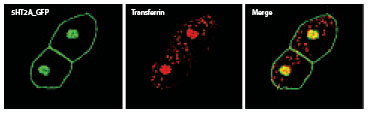
Figure 2: Antipsychotic-internalized 5-HT2A receptors are targeted to the recycling endosome. Transferrin (labeled red) co-localize with antipsychotic-internalized receptors. |
|
|
Figure 3: Serotonin present in a mouse ES colony visualized by multi-photon microscopy. High concentrations of Serotonin are present in mouse embryonic stem cells. Serotonin is distributed in a punctate pattern within these cells. Highest concentrations are in red. |

 |
Figure 4: Regulation of 5-HT1A transcripts in a conditionally-immortalized mouse hippocampal cell line. 5-HT1A mRNA transcripts expressed in P40H1 neuronal cells are down-regulated by increasing concentrations of steroids i.e. dexamethasone and hydrocortisone. Relative expression of the 5-HT1A transcript is depicted. |