
Tapomoy Bhattacharjee - Research
Future Direction

Past work
Bacterial Chemotaxis in Porous Media
Chemotactic migration of bacteria—their ability to direct multicellular motion along chemical gradients—is central to processes in agriculture, the environment, and medicine. However, studies are typically performed in bulk liquid, despite the fact that most bacteria inhabit heterogeneous porous media such as soils, sediments, and biological gels. By using direct visualization and 3D bioprinting, we find that cellular chemotaxis drives collective migration while confinement in a porous medium fundamentally alters chemotactic migration in two ways. First, cells bias their motion through a different primary mechanism in confinement than in bulk liquid. Second, confinement markedly alters the dynamics and morphology of the migrating population—features that can be described by a continuum model, but only when standard motility parameters are substantially altered from their bulk liquid values. Our work thus provides a framework to predict and control the migration of bacteria, and active matter in general, in heterogeneous environments. Read more about this work here.

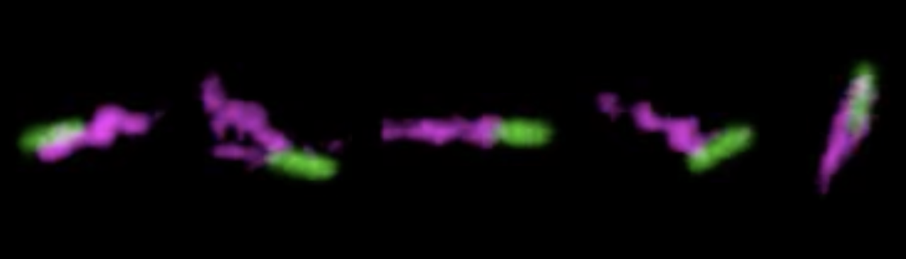
Bacterial Dynamics in 3D Porous Media
While bacterial motility is well-studied for motion on flat surfaces or in unconfined liquid media, most bacteria are found in heterogeneous porous media, such as biological gels and tissues, soils, sediments, and subsurface formations. Understanding how confinement alters bacterial motility is therefore critical to model the progression of infections, apply beneficial bacteria for drug delivery, and bioremediation. Unconfined bacteria move via runs and tumbles, leading to random walk-like motion; in a porous medium, previous research has assumed bacteria still move via runs and tumbles, but with a reduced diffusivity due to collisions with obstacles. However, this assumption has never been directly tested due to the inability to visualize processes in opaque 3D media. Here, we directly visualize the motion of single E. coli cells inside a model 3D porous medium, having controlled pore structure. By analyzing the individual cell trajectories, we find that the bacteria do not move via a run and tumble process, but instead via intermittent hops and traps reminiscent of thermally-activated transport in disordered media. We will present how bacterial motility depends sensitively on pore-scale confinement. Our findings overturn standard assumptions made in the field and provide guidance for the development of more accurate macroscopic models of bacterial motion. Our recent work can be found in Nature Communications and Soft Matter.
Jammed Microgels as 3D Cell Growth Media
Cells grown on plates differ dramatically from cells in vivo or in 3D culture in terms of cell shape, structure, motion, and mechanical behavior. These physical properties of cells in 3D are, however, far less explored. By contrast, in terms of molecular biology, it is well known that gene expression profiles of cells grown in monolayers are anticorrelated with those of cells grown in 3D culture or xenograft animal models, whereas, our traditional approach towards cell biology majorly depends on the monolayer cell culture on 2D plates. Thus, to bridge this major gap between 2D in vitro culture and 3D in vivo biology we have created a combined 3D bioprinting and culture platform by directly packing microgel pre-swelled in liquid cell growth media. Our detailed work has been published in ACS Biomaterials Science and Engineering.
We have also explored the motility of T cells in systems of jammed microgel growth media prepared with different pore sizes, finding timescale, and length-scale dependent dynamics that are correlated with the pore-space between the microgels. Our observations of cell motion through jammed microgels demonstrate how T cells navigate porous environments and provide guidance for a multitude of future investigations in unexplored territory. To find out more, please see our work published in the Journal of Physics D.


Jammed Microgels for Soft Matter Manufacturing
One of the major challenges of manufacturing soft delicate structures is to arrange them in complex 3D shapes. We overcome this constraint by using a packed system of polyelectrolyte hydrogel particles as a sacrificial support material in which we create soft delicate structures of cells, colloids, hydrogels, and elastomers with absolute high precision. We leverage the unique rheological properties of jammed microgels (low yield stress, short thixotropic time, and spontaneous reflow after yielding) to 3D print soft matter structures. Detailed work can be found in our recent work in Science Advances, MRS Bulletin and Science Advances.